


News & Events
News & Events
News
October 01, 2021
Cells act in unison when next to each other
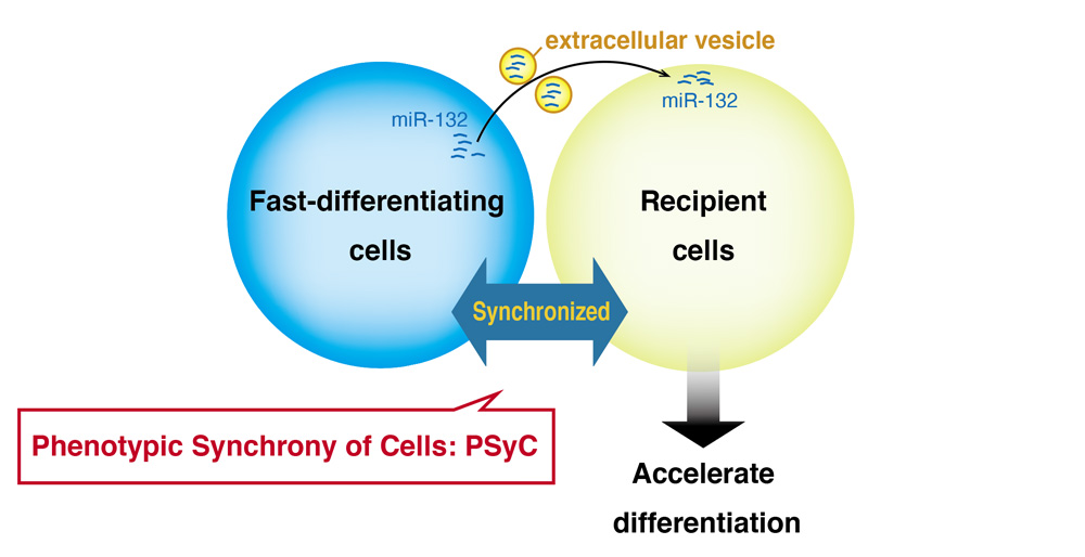
Much like an ant colony or even people in an office, cells in the body must work together to accomplish their tasks. In all cases, this cooperation depends on communication. Ants do it by smell, people by sound, and, as shown in a new study by CiRA researchers, cells do it by passing vesicles through a system called phenotypic synchrony of cells, or PSyC.
All our bodies begin as a single cell, the embryo. That cell divides, and, with time, different subsets of cells begin to form different tissues and organs. For this to happen, the cells, especially those near one another, must regularly communicate in order coordinate their activity.
"Cells must differentiate together to form normal tissues and organs," explained CiRA Professor Jun Yamashita, who led the study. "We know the signaling pathways and gene networks for individual cells, but we don't know much about how the cells synchronize."
The embryonic stem cell represents one of the earliest types of cells during embryo growth and is commonly used to study the very first steps for the development of any part of the body. To discover PSyC, the research team used a special type of embryonic stem cell that they had designed.
"We made embryonic stem cells that have protein kinase A constitutively active. This accelerates the differentiation of the cells," said Tomohiro Minakawa, who proposed the project and conducted most of the experiments.
Typically, a group of embryonic stem cells will show synchronized developmental patterns. However, because his protein kinase A embryonic stem cells show accelerated differentiation, Minakawa and his colleagues expected mixing these embryonic stem cells with regular embryonic stem cells would disrupt this synchronicity and result in two groups with different differentiation rates.
That, however, was not what they observed. While there initially was a difference in the differentiation rate of the two types of cells, the normal embryonic stem cells eventually caught up to their protein kinase A counterparts. This catching up is what the study calls PSyC.
"PSyC was only seen if the cells were in close proximity. But if we put some distance between them, the effect was lost," noted Minakawa. "This suggested the cells in close proximity were communicating by passing vesicles between each other to perform PSyC."
To exchange chemicals and molecules between one another, cells will usually enclose the content inside an extracellular vesicle, much like putting a letter into an envelope. The extracellular vesicle is then released from the cell and binds to or is absorbed by another. Then, the extracellular vesicle opens to release its package inside the second cell.
Further analysis revealed that among the many types of biomolecules passed by extracellular vesicles was microRNA-132. This exchange induced the embryonic stem cells to mesoderm cells by PSyC. To prove this point, the researchers delivered microRNA-132 inside nanoparticles to the cells, finding the same effect.
Yamashita noted that while their function in cell communication was not new, the discovery that extracellular vesicles synchronize cell activity was.
"We already knew that extracellular vesicles are used for cell-to-cell communications. But PSyC shows that they can be used to synchronize cells for cell fate and differentiation," he said.
Paper Details
- Journal: Journal of Extracellular Vesicles
- Title: Extracellular vesicles synchronize cellular phenotypes of differentiating cells
- Authors: Tomohiro Minakawa1, Tetsuya Matoba2, Fumiyoshi Ishidate3, Takahiro K. Fujiwara3,
Sho Takehana4, Yasuhiko Tabata4, and Jun K. Yamashita1 - Author Affiliations:
- Center for iPS Cell Research and Application (CiRA), Kyoto University, Kyoto, Japan
- Department of Cardiovascular Medicine, Kyushu University, Fukuoka, Japan
- iCeMS Analysis Centre, Institute for Integrate Cell-Material Sciences (WPI-iCeMS), Kyoto University, Kyoto, Japan
- Laboratory of Biomaterials, Institute for Frontier Life and Medical Sciences, Kyoto University, Kyoto, Japan


