


News & Events
News & Events
News
December 02, 2024
Seeing is believing: Leveraging defining morphological features for accurate selection of brain organoids
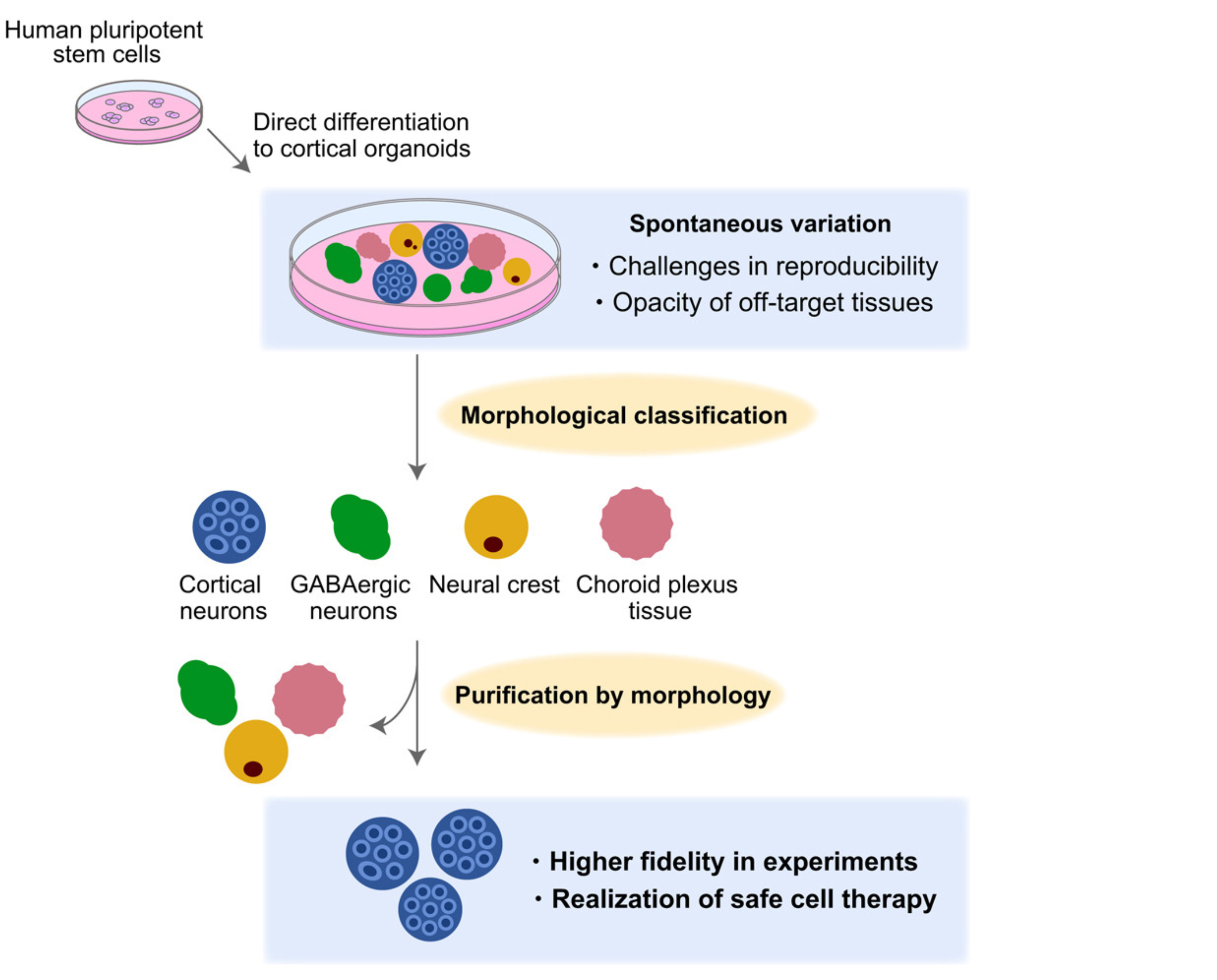
Organoids, or self-organizing miniature organs, represent a powerful new tool to study organogenesis or model diseases and are considered a means to generate cells and tissues for cell transplantation therapies. However, due to the stochastic self-organizing nature of organoids, they are highly variable and often produce non-target cell types, even with advanced induction protocols. Hence, there is a great demand for new ways to evaluate organoids in a non-destructive manner to select the most appropriate ones for transplantation.
As an ongoing effort to leverage organoid technology to produce transplantable tissues to replace damaged cells in affected brain regions following stroke or traumatic injuries, the researchers sought to develop a morphology-based evaluation system to select cerebral cortical organoids with the desired cell types for possible transplantation. From independent batches of cerebral cortical organoids, the research team classified the resulting organoids into seven types, each displaying defining morphological features, for gene expression profiling at the single-cell level by single-cell RNA sequencing (scRNA-seq). Altogether, nine groups of cerebral cortical organoids, including two groups with mixed morphological characteristics, were examined.
Through scRNA-seq analysis, the identity of individual cells from each organoid group was annotated based on marker gene expression. Various neuronal subtypes, including cortical and GABAergic neurons, and non-neuronal cells, such as fibroblasts and endothelial cells, were identified. Based on this information, the research team gained insights into the cellular compositions of different organoid types. For example, whereas organoids with rosette-like concentric layered structures were comprised primarily of cortical neurons, those with low transparency and no clear internal structures predominantly contained GABAergic neurons.
Next, the researchers validated the observed correlation between organoid morphology and cellular composition by examining the similarity in global gene expression as determined by RNA microarray analysis among independent organoids within each organoid type. In addition, a detailed analysis of organoid types based on cell type-specific marker genes and proteins showed such correlations to be reproducible across different cell lines used for organoid generation.
Finally, due to the research team's interest in using cerebral cortical organoids for cell replacement therapies in the future, their focus was on organoids with rosette-like concentric layered structures because they mostly contain cortical neurons. As such, they selected organoids with this morphological characteristic from three independent batches of cerebral cortical organoids for scRNA-seq analysis. All samples showed similar gene expression profiles, thus demonstrating remarkable reproducibility based on morphological features alone and the ability to pick desired cerebral cortical organoids correctly in a non-destructive manner. Notably, non-target cell types were not detected in the selected organoids, thus giving confidence that only desired cell types would be transplanted.
Through this work, the research demonstrated the feasibility of using simple visual information to accurately select cerebral cortical organoids that could potentially be used as a cell source of cell replacement therapy. This simple classification method is readily adaptable and could dramatically enhance the efficacy and safety of transplants.
Paper Details
- Journal: Stem Cell Reports
- Title: Validation of non-destructive morphology-based selection of cerebral cortical organoids by paired morphological and single-cell RNA-seq analyses
- Authors:
Megumi Ikeda1,2, Daisuke Doi1, Hayao Ebise2, Yuki Ozaki1, Misaki Fujii1, Tetsuhiro Kikuchi1, Kenji Yoshida2, Jun Takahashi1,*
*: Corresponding author - Author Affiliations:
- Center for iPS Cell Research and Application, Kyoto University
- Regenerative & Cellular Medicine Kobe Center, Sumitomo Pharma Co., Ltd.


