


News & Events
News & Events
News
May 19, 2026
Visualizing Early Myelin Formation Using a Nanofiber-Based Platform: A Human Cell-Based System as a Novel MPS (Microphysiological Systems) Tool
Oligodendrocytes are glial cells that wrap axons with myelin, an insulating structure essential for efficient signal transmission in the central nervous system. Disruption of oligodendrocyte function and myelin integrity is implicated in a wide range of neurological conditions, including multiple sclerosis, leukodystrophies, neurodegenerative diseases, and drug-induced leukoencephalopathies. Despite their importance, experimental models that reliably reproduce human oligodendrocyte behavior have remained limited, largely relying on rodent systems that differ substantially from humans in white matter structure, gene expression, and developmental timing. These species differences have contributed to poor translational success in therapeutic development. This platform provides a new tool to study white matter biology and evaluate compounds that modulate early myelination processes, and holds significant value as an alternative to animal-based testing as a microphysiological systems (MPS).
In this study, the research team established a robust and reproducible method to rapidly generate human oligodendrocytes from iPS cells using inducible expression of key transcription factors. To recreate physical features of axons without the complexity of neuron co-culture, differentiated oligodendrocytes were cultured on aligned nanofibers with diameters comparable to human axons. This engineered scaffold provides a defined microenvironment in which oligodendrocyte processes can extend, interact with fiber-like structures, and initiate ensheathment.
Using ultrastructural analyses, live-cell imaging, and transcriptomics, the researchers showed that oligodendrocytes dynamically probe the nanofibers and form wrapping structures reminiscent of early axonal ensheathment. Rather than simply promoting terminal maturation, nanofiber culture induced a specific molecular program characterized by enhanced lipid metabolism, cell adhesion, and extracellular matrix organization--pathways essential for membrane expansion and structural organization during myelination. Importantly, conventional differentiation markers alone were insufficient to capture these early events.
A central advance of the platform is the use of Claudin-11, a myelin-specific adhesion molecule, as a quantitative and spatial readout of structural alignment during ensheathment. Claudin-11 signals became highly oriented along nanofibers, reflecting polarized membrane organization that precedes compact myelin formation. By combining Claudin-11 alignment with image-based analysis, the researchers established a sensitive assay to evaluate how compounds influence oligodendrocyte structural organization.
As proof of concept, the platform successfully detected the effects of known enhancers and inhibitors of ensheathment, as well as direct oligodendrocyte toxins associated with clinical white matter injury. These results demonstrate that the system can function both as a screening tool for therapeutic candidates that promote myelin initiation and as an assay to identify compounds with potential white matter toxicity.
While the model focuses on the initial phase of ensheathment rather than fully compacted myelin, this stage represents a critical and previously difficult-to-evaluate step in myelination. By providing a human-specific, experimentally accessible system with quantitative structural readouts, this platform offers a powerful foundation for studying oligodendrocyte pathology, understanding white matter vulnerability, and accelerating the development and safety evaluation of drugs targeting myelin-related disorders.
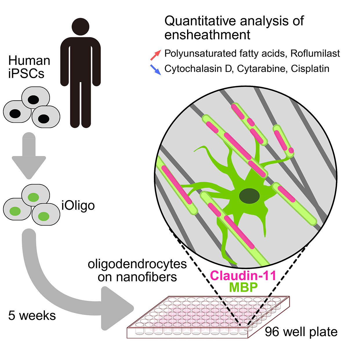
Graphical abstract
Paper Details
- Journal: Stem Cell Reports
- Title: Nanofiber-based platform for quantitative analysis of human oligodendrocyte ensheathment with pharmacological perturbations
- Authors: Satoshi Morita1,2,3, Takayuki Kondo1,2,4, Keiko Imamura1,2,4, Yukako Sagara2, Kayoko Tsukita1, Hisanori Tokuda3, Yoshihisa Kaneda3, Takayuki Izumo3, Yoshihiro Nakao3, Haruhisa Inoue1,2,4,*
*: Corresponding author - Author Affiliations:
- Center for iPS Cell Research and Application (CiRA), Kyoto University
- iPSC-based Drug Discovery and Development Team, RIKEN BioResource Research Center (BRC)
- Institute for Science of Life, Suntory Wellness Ltd.
- Medical-risk Avoidance based on iPS Cells Team, RIKEN Center for Advanced Intelligence Project (AIP)


