






News & Events
News & Events
News
July 23, 2026
From Growth to Function: PRDM16 Helps Human Heart Cells Decide When to Mature
Understanding how heart muscle cells stop dividing and acquire the characteristics needed to sustain lifelong cardiac function remains one of the greatest challenges in cardiovascular biology. Leveraging human induced pluripotent stem (iPS) cell technology, a team led by Associate Professor Yoshinori Yoshida (Department of Clinical Application, CiRA, Kyoto University) and Associate Professor Antonio Lucena-Cacace (WPI-PRIMe, The University of Osaka) has identified PRDM16 as an important regulator governing the balance between proliferation and maturation in human iPSC-derived cardiomyocytes.
The study reveals that PRDM16 acts as a developmental "rheostat": low levels permit cardiomyocytes to retain proliferative competence, whereas higher levels facilitate the acquisition of structural, metabolic, and functional characteristics associated with more mature heart cells. The findings provide a framework for improving regenerative strategies and generating higher-quality cardiac tissues for disease modeling and drug discovery.
Successful cardiac regeneration requires overcoming a fundamental biological dilemma. During embryonic development, cardiomyocytes proliferate extensively to build the heart. Shortly after birth, however, these cells progressively withdraw from the cell cycle and adopt specialized functions that support lifelong contraction. While this maturation process is essential for cardiac performance, it severely restricts the regenerative capacity of the adult human heart following injury.
Similarly, cardiomyocytes generated from iPS cells remain relatively immature, limiting their utility for translational applications. Understanding the molecular signals coordinating the transition from proliferation to maturation has therefore become a major objective in regenerative medicine.
Using fluorescent cell-cycle reporter systems, transcriptomic analyses, engineered heart tissues, and gain- and loss-of-function approaches, the team demonstrated that PRDM16 occupies a central position in this developmental transition.
"When we reduced PRDM16 levels, cardiomyocytes regained aspects of proliferative competence that are normally lost during maturation," explains Kanae Tani, researcher and first author of the study. "At the same time, these cells struggled to acquire adult-like characteristics, suggesting that PRDM16 is involved in coordinating the trade-off between growth and specialization."
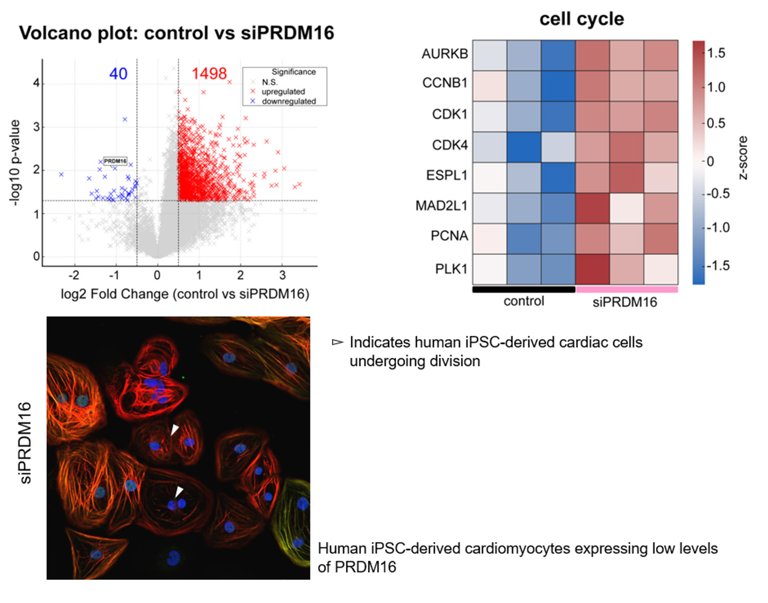
Volcano plot: Volcano plot of differentially expressed genes between control and siPRDM16 groups.
Heat map: Heatmap of selected cell cycle regulators, demonstrating consistent upregulation in siPRDM16 hiPSC-CMs compared with controls.
Immunocytochemistry analysis: Immunofluorescence staining of hiPSC-CMs for TNNI1 (red; immature isoform) and TNNI3 (green; mature isoform) under siPRDM16 conditions. Nuclei are counterstained with Hoechst (blue).
Indeed, PRDM16-deficient cardiomyocytes displayed elevated expression of proliferative regulators, including CDK1 and phospho-AKT, increased cell-cycle activity, and impaired acquisition of mature sarcomeric organization and mitochondrial function. Engineered heart tissues generated from these cells also exhibited diminished contractile performance.
Conversely, moderate overexpression of PRDM16 suppressed proliferation while promoting hallmarks of cardiomyocyte maturation, including cellular hypertrophy, increased expression of adult cardiac proteins such as TNNI3, enhanced oxidative metabolism, and reduced spontaneous beating frequency.
"Our findings suggest that PRDM16 functions as a molecular checkpoint guiding cardiomyocytes toward functional competence," says Yoshinori Yoshida, who supervised the study. "Understanding how this balance is achieved may ultimately allow us to generate cardiac tissues that are both physiologically relevant and clinically useful."
The study also raises the possibility that temporal manipulation of PRDM16 could one day contribute to regenerative strategies.
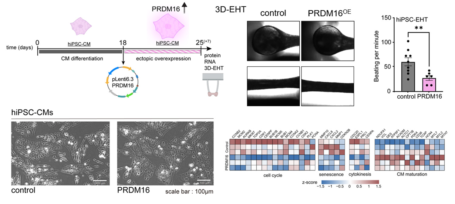
Scheme: Experimental schematic showing differentiation of hiPSCs into cardiomyocytes followed by ectopic overexpression of PRDM16 using a lentiviral vector (pLenti6.3-PRDM16).
Brightfield images: Representative images of engineered heart tissues (3D-EHTs) generated from control and PRDM16 OE.
Graph: Beating frequency of hiPSC-derived engineered heart tissues generated from control and PRDM16-overexpressing cardiomyocytes.
Microscopic images: Representative phase contrast images of hiPSC-CM monolayers under control and PRDM16-overexpressing conditions.
Heatmap: Heatmap showing cell cycle, senescence, cytokinesis and maturation genes expression in PRDM16-overexpressing hiPSC-CMs relative to control.
"One of the long-standing goals in cardiac biology has been to recover proliferative potential without permanently compromising maturation," notes Antonio Lucena-Cacace, co-corresponding author of the study. "PRDM16 appears to occupy an interesting position within this continuum. While mature cardiomyocytes require sufficient PRDM16 activity to acquire specialized functions, transient modulation of this pathway may provide opportunities to enhance regenerative responses or improve the quality of stem cell-derived cardiac models."
The researchers emphasize that further studies will be required to identify the direct genomic targets of PRDM16 and determine how its regulatory activity changes throughout cardiac development. Nevertheless, the present findings establish PRDM16 as a previously underappreciated determinant of human cardiomyocyte biology.
Published in Stem Cell Reports, this work places PRDM16 among a growing number of developmental regulators being explored to optimize iPS cell-derived cardiac systems. By clarifying how human cardiomyocytes transit between proliferation and maturation, the study advances efforts to engineer more faithful models of the human heart and develop future regenerative therapies.
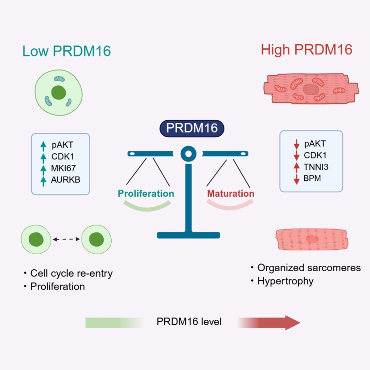
Graphical abstract
Paper Details
- Journal: Stem Cell Reports
- Title: PRDM16 Modulates Aspects of Cell-Cycle Dynamics and Maturation in Human iPSC-Derived Cardiomyocytes
- Authors: Kanae Tani1, Yasuko Matsumura1, Misato Nishikawa1, Megumi Narita1, Amanda Putri-Elvandari1, Antonio Lucena-Cacace1,2*, Yoshinori Yoshida1*
*: These authors contributed as co-corresponding authors. - Author Affiliations:
- Center for iPS Cell Research and Application (CiRA), Kyoto University
- Premium Research Institute for Human Metaverse Medicine (WPI-PRIMe), The University of Osaka