


News & Events
News & Events
News
July 11, 2025
Revealing how senescent cells shape aging at the single-cell level
Cellular senescence is a state of irreversible cell cycle arrest triggered by various stressors, including oncogene activation, DNA damage, and oxidative stress. While senescence serves as a protective mechanism against tumorigenesis and contributes to tissue repair, its accumulation over time is associated with aging and chronic diseases. Despite its importance, the physiological role of senescence in vivo has remained poorly understood due to the lack of reliable models and the difficulty of identifying senescent cells within complex tissue environments.
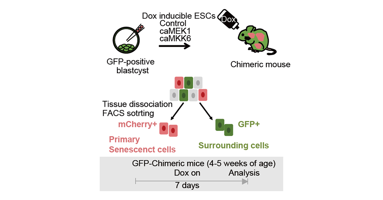
To address this, the researchers engineered two mouse models that allow inducible expression of constitutively active MEK1 (caMEK1) or MKK6 (caMKK6), which activate the ERK and p38 MAPK pathways, respectively—both known to trigger senescence in vitro. These models were combined with a dual-color labeling system, enabling the distinction between primary senescent cells (red fluorescence) and surrounding secondary senescent cells (green fluorescence). This setup allowed the team to analyze gene expression at single-cell resolution and trace the spread of senescence within tissues.
Both caMEK1 and caMKK6 induced hallmark features of senescence in liver and colon tissues, including elevated p21 expression, DNA damage responses, and the senescence-associated secretory phenotype (SASP). Transcriptomic analyses revealed that senescent cells exhibit highly diverse gene expression profiles depending on tissue type, senescence inducer, and even spatial location within the same tissue. Notably, SASP factors such as IL-1β, primarily secreted by macrophages, were shown to induce secondary senescence in neighboring cells. Notch signaling was also implicated in both primary and secondary senescence, contributing to lateral induction through cell-to-cell communication and reinforcing the importance of intercellular signaling in shaping the senescent microenvironment.
A particularly striking finding was that senescence disrupted liver zonation—a spatial organization essential for liver metabolic function—especially in caMEK1-induced models. This disruption led to impaired expression of genes involved in metabolism, mirroring changes observed in naturally aged tissues. Comparative analyses confirmed that the transcriptomic profiles of senescent cells in these models closely resembled those found in aged mice and humans, suggesting that senescence plays a causal role in age-related tissue dysfunction.
This study provides the first comprehensive in vivo characterization of primary and secondary senescent cells at single-cell resolution. By offering unprecedented insights into how senescent cells function within living tissues and affect surrounding cells, the research lays a foundation for future studies and therapeutic strategies targeting senescence. The caMEK1 and caMKK6 mouse models represent powerful tools for advancing our understanding of aging at the organismal level.
Paper Details
- Journal: Nature Aging
- Title: Characterizing primary and secondary senescence in vivo
- Authors:
Yuko Sogabe1,2, Hirofumi Shibata1,7, Mio Kabata1, Akito Tanaka1, Kanae Mitsunaga1, Kazunori Sunadome1,3, May Nakajima-Koyama1, Michitada Hirano4, Eisuke Nishida5, Knut Woltjen1, Hiroshi Seno2, Yasuhiro Yamada4*, Takuya Yamamoto1,3,6*
*: Corresponding author - Author Affiliations:
- Department of Life Science Frontiers, Center for iPS Cell Research and Application (CiRA),
Kyoto University - Department of Gastroenterology and Hepatology, Graduate School of Medicine, Kyoto University
- Institute for the Advanced Study of Human Biology (WPI-ASHBi), Kyoto University
- Department of Molecular Pathology, Graduate School of Medicine, The University of Tokyo
- RIKEN Center for Biosystems Dynamics Research (BDR)
- Medical-risk Avoidance Based on iPS Cells Team, RIKEN Center for Advanced Intelligence Project (AIP)
- Present address: Department of Otolaryngology, Gifu University Graduate School of Medicine
- Department of Life Science Frontiers, Center for iPS Cell Research and Application (CiRA),


